Years ago, I got into a heated exchange with an Italian dentist on social media. I cringe when I reflect on it now.
The dentist shared that he had a patient with severe obstructive sleep apnea (OSA) who was unable to use his CPAP machine. Fitting a custom oral appliance, the dentist reduced the man’s apnea-hypopnea index (AHI) from the 70s to only nine events per hour. Instead of applauding a good outcome and the improved quality of life the patient was experiencing, I complained, “Obstructive sleep apnea isn’t properly treated until the AHI is under five events per hour.”
The exasperated dentist calmly pointed out that the patient had abandoned his CPAP therapy and had left his OSA entirely untreated before he got his oral appliance.
“Isn’t an AHI of nine with an oral appliance better than an AHI in the 70s with no treatment?” he asked.
Since this exchange, I have been ruminating on this question. In that time, I have recorded more than 140 episodes of my podcast, “Sleep Apnea Stories,” interviewing people living with sleep apnea about their treatment choices as well as experts from different specialties. I have learned so much about the broad spectrum of experiences in our sleep apnea community and the reasoning behind individual treatment choices.
I can understand the frustration of board-certified sleep specialists who prescribe CPAP therapy to their patients, only to see it abandoned. I am a huge advocate of more support and resources for new CPAP users struggling to adapt to their therapy. Early intervention with practical troubleshooting and empathy can be all that’s needed to take someone from giving up on their therapy to successful adherence. This could be a group CPAP therapy clinic for new users to get support in person, or an online coaching model similar to what companies like Lofta and BetterNight provide.
The very best sleep clinics offering superb support to new CPAP users still have a significant number of people who either never start CPAP therapy or abandon it over time. For those people who are leaving their OSA entirely untreated, we need a new attitude of pragmatism. Arguing that CPAP therapy offers the best results in people who use it isn’t helping the group who won’t or can’t use their machine.
The great news for patients is that the range of viable treatment options for OSA is expanding. The GLP-1 tirzepatide is already available as Zepbound, an injectable medication for people with both OSA and obesity. The oral pill version orforglipron has recently reported positive phase 3 trial results. Also in the “coming soon” category is Apnimed’s AD109, a once nightly oral pill that works to maintain upper airway muscle tone during sleep. The successful phase 3 trials for AD109 open up a whole new frontier of using pharmacotherapy to target the cause of OSA.
The dental sleep medicine community, not to be outdone, has been working hard on appliances with integrated SpO2 sensors. These oral appliances will enable the sharing of data, including wear time, oxygen desaturation index, pulse rate, and more, not only with patients but also directly with doctors.
Ear, nose, and throat surgeons have more techniques at their disposal than ever too. Top surgeons continue to refine throat surgeries to include new techniques like transoral robotic surgery. Hypoglossal nerve stimulation implants are evolving with the new Inspire V system with quicker surgery time and a Bluetooth patient remote. Nxyoah’s Genio neurostimulator just earned FDA clearance.
With so many treatment options currently available, and on the horizon, it is time we adopt a more pragmatic approach to considering a person’s OSA “well-treated.” Offering a treatment other than CPAP to lessen the severity of the OSA is far better for quality of life and health outcomes than no treatment at all.
As a patient advocate, I feel strongly that every person with an OSA diagnosis should be educated on and offered all the treatment options that could be useful to them.
I have, in short, changed my tune and now agree with the Italian dentist I argued with. I wish I could remember his name so I could apologize directly. Reducing the severity of obstructive sleep apnea and improving the quality of life for each individual is a worthy goal, even without hitting fewer than five events per hour.
A study published in Nature Reviews Neuroscience by an international team including the Woolcock’s Dr. Rick Wassing examined research into sleep disorders over more than two decades to prove a good night’s sleep is the perfect remedy for emotional distress.
Nothing we haven’t known forever, some would argue, but Dr. Wassing who has spent the past two years on the project says there’s much more to it than that.
“What we have done with this study is explain why. We looked at studies in neurobiology, neurochemistry and clinical psychology to get a real understanding of the mechanisms underlying how sleep helps us to deal with our emotional memories.”
What the team of researchers believe after aggregating more than 20 years’ of scientific knowledge is that the way certain neurochemicals (for example, serotonin and noradrenaline) are regulated during sleep is crucial for the processing of emotional memories and our long-term mental health.
Chemistry and circuitry
Serotonin is involved in many, if not almost all, aspects of learning emotional experiences. It helps us assess and understand the world around us. Noradrenaline is all about “fight or flight”—it allows us to assess and respond to danger. Both are turned off during rapid eye movement (REM) sleep and that creates this “really beautiful opportunity for the brain to engage in processes that are otherwise not doable when we are awake,” explains Dr. Wassing.
There are two main ways we process emotional memory during sleep, he says, and they involve the brain’s hippocampus and amygdala.
Our brains store what we learn each day. This learning is governed by the hippocampus aggregating and cataloging this new information into the “novelty” memory store as we process it. At the same time, if that new experience is emotional, the amygdala is very active and coupled with the autonomic nervous system—think racing heart, knots in your stomach, skin crawling.
During REM sleep, our brains reactivate these new memories. It is as if the brain replays a summary of what had happened when we experienced the memory. But during REM sleep, when the noradrenergic and serotonergic systems are turned off, these memories can be moved into the “familiar” storage without experiencing the physical “fight or flight” response. That can’t happen while we’re awake or—as is the case for people with sleep disorders—when we don’t get consistent blocks of REM sleep.
Shining a light on the brain
Much of what we now know about the way information is processed by the brain comes from the relatively new field of optogenetics which is used to activate or inhibit very specific cell types in a neuronal network. This has allowed researchers to see what cell types and brain regions are involved in encoding emotional memories.
According to Dr. Wassing, it has meant real breakthroughs in terms of our understanding of brain circuitry and neurobiology.
It’s all well and good, he says, to look at neurons and receptors and circuits, but the researchers also assessed clinical psychology studies and found that their findings, especially relating to disconnecting amygdala reactivity and shutting down the autonomic nervous system, were corroborated.
“All three levels of neuroscience align to produce the same conclusion, that the way the brain functions during REM sleep is important for processing emotional memories.”
Making ‘good sleepers’
So, where to now? “We know that with insomnia or other sleep disorders where people wake up from sleep a lot, we see an increased risk of developing mental health problems. Our hypothesis would be that that these awakenings from sleep lead to the fact that the noradrenergic system is not shut down for long periods of time (in fact, they might actually show enhanced activity) and that’s why these people might not be able to regulate emotional memories.”
“The solution is to try to get a good night’s sleep, yes, but the problem is how then do we do that? We know that two out of three people with insomnia benefit from cognitive behavioral therapy for insomnia (CBTI) but that is mostly based on subjective ratings. There’s less evidence on objective sleep measures. The insomnia patient after CBTI is not necessarily a good sleeping individual, they still have some sleep disturbances but CBTI is enabling them to better deal with them.”
“We need to critically think more about the mechanisms that regulate sleep. It’s very hard to target one system because sleep is very dynamic—the noradrenergic system shuts down during REM sleep, but it actually needs to be active during non-REM sleep so you can’t just turn it off for the entire sleep cycle.”
“We need really creative ideas about how to design an intervention or a drug that can target these dynamics that happen during sleep and enable those systems to renormalize. We need to be targeting objective sleep and making people with insomnia good sleepers again.”
A cross-sectional study utilizing self-reports from Chinese college students correlated mobile phone use with insomnia, bolstering previous research that made similar conclusions.
A study recently published in Frontiers Public Healthreinforced the known correlation between mobile phone addiction and insomnia, while additionally suggesting that increasing one’s physical activity could mitigate this negative impact.
Man Endlessly Scrolling in Bed | image credit: Louis-Photo – stock.adobe.com
During the COVID-19 pandemic, the use of social media was seen as a benefit both for providing social support during large-scale isolation and disseminating information on public health. However, as the authors of the current study mentioned, the negative consequences associated with mobile phone use—such as its documented influence on insomnia—should not be ignored. The authors added that investigations into the effects of mobile phone use during the pandemic have largely focused on the mental health of adolescents while the outcomes related to insomnia have been unknown.
To address this gap in knowledge, researchers conducted a study to investigate what underlying factors influence the relationship between insomnia and mobile phone addiction in college students, considering the mediating role of social anxiety and the moderating role of physical activity. This study was conducted in China, which endured the first outbreak of COVID-19, and surveyed Chinese college students.
This study was conducted with an online questionnaire, which was completed by 301 college students. A student’s degree of mobile phone addiction was measured with the Chinese iteration of the Mobile Phone Addiction Tendency Scale (MPATS), where higher scores indicate more intense addiction. The Chinese iteration of the Social Phobia Inventory (SPIN) was used to measure social anxiety, and higher scores indicate higher levels of social anxiety. Physical activity levels were measured with the Physical Activity Rating Scale (PARS-3) and insomnia by the Insomnia Severity Index where, similarly in both, higher scores indicate higher levels of activity and more severe insomnia.
Their results showed that mobile phone addiction had a positive association with social anxiety (P < .001) and that social anxiety was positively associated with insomnia (P < .01). Overall, the correlation between mobile phone addiction and insomnia was found to be significant (P < .001). These findings suggested that social anxiety could be a mediator of this relationship because the indirect effect between them was also found to be significant (indirect effect = 0.03; SE = 0.01; 95% Boot CI, 0.01–0.06).
The self-reports indicated that more physical activity could reduce both feelings of social anxiety as well as feelings of addiction to one’s mobile phone. This moderating role of physical activity on the relationship between insomnia and mobile phone addiction was found to be significant (P < .05). The authors went on to comment that physical activity has previously demonstrated to be beneficial for the management of stress and loneliness, and can improve one’s mental health. The authors’ findings were consistent with prior reports in this area.
“But it is worth noting that social anxiety could only partially account for the relationship between mobile phone addiction and insomnia; thus other key latent indicators, such as interpersonal distress, should also considered when examining the mediation role between them afterwards,” they added.
The limitations of the study include the inability to establish causal relationships due to the cross-sectional design, the risk of recall bias that comes with self-reported data, and the use of convenient sampling which resulted in a moderate sample size. Additionally, the exclusive focus on college students during a specific pandemic period may limit the generalizability of the findings.
“In summary, our research results indicate that increasing physical activity and reducing social anxiety can improve insomnia symptoms among college students,” the authors concluded. “To prevent future problematic behaviors among college students, it is necessary to adopt proactive health behaviors, such as engaging in physical activity, to counteract or avoid subsequent passive impacts.”
Reference
Wang J, Xu X, Zuo L, Wang H, Yang G. Mobile phone addiction and insomnia among college students in China during the COVID-19 pandemic: a moderated mediation model. Front Public Health. 2024;12:1338526. doi:10.3389/fpubh.2024.1338526
Comments Off on Why people should prioritize sleep quality over quantity.
by SadofNE
By Analisa Novak – February 19, 2024 / 11:59 AM EST / CBS News
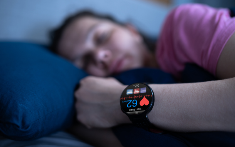
When it comes to maintaining heart health, it’s not just how long you sleep — it’s how well you sleep that matters equally, if not more, said Dr. Shelby Harris, a behavioral sleep psychologist and clinical associate professor at Albert Einstein College of Medicine.
“Poor quality sleep really can influence our heart health as well,” Harris told “CBS Mornings” in an interview during American Heart Month.
Harris said the body’s balance of ghrelin and leptin, hormones that regulate hunger, is also disrupted by poor sleep, leading to increased consumption of high-sugar and high-fat foods.
Sleep disorders like sleep apnea and insomnia are closely linked to heart health. Sleep apnea, characterized by snoring and breathing pauses, affects both men and women, though women are evaluated less frequently for it.
Strategies for improving sleep quality include limiting alcohol and caffeine intake, reducing screen time before bed and managing how much liquid you drink before bed.
“Once you have better quality then we try to work on the quantity of sleep possible,” Harris said.
For those struggling to achieve a longer sleep duration, getting a good quality amount of sleep but shorter is ideal when you first are trying to tackle this goal.
“For some people, I might have them go to bed later and then I might slowly have them go to bed earlier over time as opposed to that shifting back and forth. Because that can create a lot of problems for people as well,” she said.
Harris said that sleeping pills and aids are not ideal for most people, but said cognitive-behavioral therapy for insomnia may offer an alternative solution to medication.
She said most people try four to eight sessions of the therapy and if that doesn’t work, then she would recommend sleeping aids.
“You work on not just the hygiene but you work on the timing of sleep. … We work on thoughts about sleep, a lot of people put pressure on themselves to sleep and they worry about what’s going to happen if they don’t sleep and so we work on that aspect,” she said.
Harris said dietary choices also play a role in a good night’s sleep. She said people should avoid consuming large or heavy meals, such as a big dinner, before going to bed.
Instead, opting for a small, light snack that includes a mix of protein and carbohydrates can be beneficial. “That’s a really good mix to help you throughout the night so you don’t wake up hungry, which a lot of people do as well,” said Harris.
Dementia is also associated with abnormalities in the brain’s white matter that are hallmarks or markers of cerebrovascular diseases.
A recent observational study shows that severe obstructive sleep apnea and reduced deep sleep were independently associated with white matter abnormalities related to cerebrovascular diseases in cognitively unimpaired older adults.
These findings show that severe obstructive sleep apnea and poor sleep quality can lead to an increase in the biomarkers of cerebrovascular disease, potentially increasing the risk of cognitive decline and stroke.
Obstructive sleep apnea is the most common type of sleep-breathing disorder that affects nearly a billionTrusted Source individuals across the globe.
A recent study published in Neurology suggests that obstructive sleep apnea and a reduction in deep sleep, also known as slow-wave sleep, were independently associated with an increase in white matter abnormalities in the brain.
The white matter abnormalities assessed in the study are known markers of cerebrovascular disease and are also observed in mild cognitive impairment and Alzheimer’s disease.
The findings from this observational study thus suggest that obstructive sleep apnea and poor sleep quality could potentially lead to increased white matter abnormalities, subsequently increasing the risk of dementia and stroke.
The study’s author Dr. Diego Carvalho, a neurologist at the Mayo Clinic in Rochester, MN, told Medical News Today:
“White matter abnormalities increase with aging and may contribute to cognitive decline, dementia, and stroke. Since there is no treatment to reverse or slow them down other than risk factor prevention, it is important to understand what may contribute to their development.”
“In our study, we found that severe sleep apnea and decreased deep sleep were associated with more white matter abnormalities. Although we cannot infer a direct causal relationship with a cross-sectional study design, the findings raise the possibility that sleep interventions may prevent the progression of white matter disease. Although there is already compelling evidence that sleep apnea is involved in white matter abnormalities, the potential role of slow-wave sleep (or deep sleep) in white matter health is much less understood,” added Dr. Carvalho.
Obstructive sleep apneaTrusted Source is a sleep-breathing disorder characterized by episodes of interruption of breathing due to partial or complete blockage of the upper airway. The episodes of reduced breathing are known as hypopnea, whereas apnea refers to events involving a complete blockage of the upper airway.
The apnea-hypopnea index (AHI) describes the number of apnea and hypopnea events per hour. Specifically, obstructive sleep apnea involves at least five such episodes of apnea or hypopnea per hour.
The interruption of breathing triggers a compensatory response that leads to arousal from sleep. Thus, obstructive sleep apnea leads to sleep disturbances and an experience of feeling unrefreshed after sleep.
Several studies have shown that poor sleep quality is associated with an increased risk of cognitive decline and dementia.
The accumulation of misfolded deposits of the amyloid-beta and tau proteins is a hallmark of Alzheimer’s disease. A previous study showed a higher accumulation of the amyloid-beta protein in the brains of individuals experiencing excessive daytime sleepiness.
In contrast, a brain imaging studyTrusted Source showed that cognitively unimpaired individuals with higher tau levels in their brains were at an increased risk of obstructive sleep apnea.
These studies suggest a bidirectional relationship between sleep quality and pathological changes associated with Alzheimer’s disease.
In addition to the accumulation of misfolded proteins, individuals with dementia also show damage to neurons.
The brain tissue can be categorized into white matter and gray matter. The gray matter consists of the cell bodies of neurons, whereas the white matter consists of axons that transmit information.
Several of the axonal processes in the white matter are encased in an insulating layer called the myelin sheath. The myelin sheath gives white matter its color and allows the axons to conduct electrical impulses more rapidly and efficiently.
Individuals with dementia and mild cognitive impairment show abnormalities in the white matter. Some of these white matter abnormalities, such as white matter hyperintensitiesTrusted Source and a decline in the integrity of the white matter tract, are also markers for cerebrovascular diseases, which are diseases of the blood vessels in the brain.
White matter hyperintensities are hyperintense regions identified using MRI that represent lesions of white matter generally caused by cerebral small vessel disease.
The integrity of the white matter tract is measured in terms of fractional anisotropy using a technique called diffusion tensor imaging. These white matter abnormalities due to damage to blood vessels may contribute to cognitive decline.
Sleep disorders such as obstructive sleep apnea are also associatedTrusted Source with increased risk of cerebrovascular diseases. Thus, sleep disorders could potentially lead to cerebrovascular disease-related white matter abnormalities and increase the risk of dementia.
For instance, there is evidence from the authors’ own work showing that individuals with daytime sleepiness have elevated levels of the neurofilament light chain protein in their blood, though research evidence is contradictoryTrusted Source.
The neurofilament light chain protein is a protein associated with the myelin sheath covering the axons of neurons. Thus, elevated neurofilament light chain protein levels suggest damage to myelinated axons and, thus, white matter damage.
In the present study, the researchers examined the association between sleep quality, including the presence of obstructive sleep apnea, with white matter abnormalities in the brain of cognitively unimpaired individuals.
The new study consisted of 140 individuals participating in the Mayo Clinic Study of Aging (MCSA), a population-based cohort study that aims to characterize the prevalence and risk factors associated with mild cognitive impairment and dementia.
The study included individuals who had previously undergone a brain MRI scan and at least one polysomnography test as a part of the MCSA study.
A polysomnographic study is a sleep study that assesses multiple parameters associated with sleep, including brain waves, breathing and heart rate, and blood oxygen levels. The average duration between the brain MRI scan and the polysomnography test was 1.74 years.
The researchers aimed to only include participants who were cognitively unimpaired at the time of both the MRI and the polysomnography test. The study consisted of 90.7% of the participants who were cognitively unimpaired at the time of both assessments.
Only participants with obstructive sleep apnea were included in the study. These participants were categorized as having either mild, moderate, or severe obstructive sleep apnea on the basis of the number of episodes of apnea and hypopnea per hour.
The researchers first examined the association between sleep patterns and white matter abnormalities. Sleep can be dividedTrusted Source into the non-rapid eye movement (NREM) and rapid eye movement (REM) phases.
Furthermore, the NREM phase can be further subdivided into N1-N3 phases, with the N1 being the lightest sleep phase and N3 involving deep sleep. These phases show differences in their patterns of brain waves, eye movements, and muscle tone.
Using brain waves collected during polysomnography, the researchers found that a lower fraction of time spent in the N3 phase or slow wave sleep was associated with elevated levels of white matter damage.
This association was present after accounting for variables such as age, sex, genetic risk of Alzheimer’s disease, and cardiovascular risk factors.
In a separate analysis, the researchers looked at the association between obstructive sleep apnea severity and markers of white matter damage. They categorized patients as either having severe or mild-to-moderate obstructive sleep apnea and matched individuals from the two groups for age, sex, and N3 sleep levels for this analysis.
Individuals with severe obstructive sleep apnea showed higher white matter abnormalities than those with mild-to-moderate.
Individuals in the two groups did not show differences in cardiometabolic risk factors, but the individuals with severe obstructive sleep apnea showed higher arousal levels. This indicates the fragmentation of sleep in individuals with severe obstructive sleep apnea.
Dr. Sandra Narayanan, a board-certified vascular neurologist and neuro-interventional surgeon at Pacific Stroke & Neurovascular Center at Pacific Neuroscience Institute in Santa Monica, CA, not involved in the research told us that these findings show that, while obstructive sleep apnea is associated with cardiovascular disease, it could independently increase the risk of cerebrovascular diseases.
Dr. Narayanan said: “[Obstructive sleep apnea] is an important vascular comorbidity, as it is significantly associated with an increased risk of hypertension, cardiovascular disease, and stroke. This study demonstrates a separate association of OSA with imaging biomarkers of cerebrovascular disease.”
Some of the previous studies showing a link between sleep quality and white matter abnormalities have not controlled for cardiometabolic risk factors. These cardiometabolic factors can increase the risk of cerebrovascular conditions, such as stroke, thus potentially biasing the results.
One of the strengths of the present study was that the researchers controlled for cardiometabolic risk factors.
The authors acknowledged that their study had a few limitations. They noted that they only collected sleep data during the initial few hours of sleep.
This could have biased the data on sleep patterns. For instance, the period of REM sleep tends to increase during the night, whereas the duration of deep sleep tends to decline.
Dr. Narayanan noted: “While the imaging biomarkers of CVD noted in this study were not independently linked during the course of this study to the development of incident stroke, the presence of white matter hyperintensities is strongly associated with cognitive impairment, stroke, and death in numerous other studies.”
“Fractional anisotropy (FA) is a marker of white matter integrity, as noted in diffusion tensor imaging (DTI),“ she explained. “Decreased FA is associated with other neurodegenerative disorders such as Alzheimer’s dementia and Parkinson’s disease, but has a poor prognostic value for motor recovery following stroke.”
The authors also noted that the study had an observational design, and further studies are needed to show that obstructive sleep apnea and reduced slow-wave sleep can cause an increase in the biomarker of cerebrovascular disease.
Comments Off on Too Little Sleep Might Raise a Woman’s Odds for Diabetes
by SadofNE
Original Article | Dennis Thompson, Published in HealthDay Magazine
Key Takeaways
Women who get poor sleep might have an increased risk of diabetes
Getting just 90 minutes less sleep increased insulin resistance in women
Researchers will look at whether better sleep helps control diabetes
TUESDAY, Nov. 14, 2023 (HealthDay News) — Women who don’t get enough sleep might have an increased risk of diabetes, an effect even more pronounced in postmenopausal females, a new study finds.
Shortening sleep by just 90 minutes increased insulin resistance in women used to getting adequate sleep, researchers at Columbia University.
The findings are the first to show that even a mild sleep deficit maintained for six weeks can raise the risk of diabetes, researchers said.
“Throughout their lifespan, women face many changes in their sleep habits due to childbearing, child-rearing and menopause,” said lead researcher Marie-Pierre St-Onge, director of the Center of Excellence for Sleep and Circadian Research at Columbia University in New York City. “And more women than men have the perception they aren’t getting enough sleep.”
For this study, St-Onge and her colleagues enrolled 38 healthy women, 11 of whom had gone through menopause.
All of the women routinely slept at least seven hours each night. The recommended amount of sleep for optimal health is between seven and nine hours, researchers said, but about a third of Americans get less sleep than that.
Each of the women were asked to participate in two different phases of the study, in random order.
Women were asked to maintain their regular adequate sleep in one phase, but in the other phase they were asked to delay their bedtime by an hour and a half, shortening their total sleep to around six hours. Each phase lasted six weeks.
Curtailing sleep by 90 minutes for six weeks increased fasting insulin levels by more than 12% overall, and by 15% among premenopausal women.
Insulin resistance increased by nearly 15% overall, and by more than 20% among postmenopausal women.
Average blood sugar levels remained stable for all participants throughout the study, but researchers said the changes in insulin resistance could cause them to start rising in the long-term.
Although increased belly fat is a key driver of insulin resistance, the researchers found that the effects of sleep loss on insulin resistance were not linked to any increases in fat.
“The fact that we saw these results independent of any changes in body fat, which is a known risk factor for type 2 diabetes, speaks to the impact of mild sleep reduction on insulin-producing cells and metabolism,” St-Onge said.
Researchers will next investigate whether better sleep can improve blood sugar control and glucose metabolism.
The study was published Nov. 13 in the journal Diabetes Care.
More information
The U.S. Centers for Disease Control and Prevention has more about sleep and diabetes.
SOURCE: Columbia University, news release, Nov. 13, 2023
To provide the best experiences, we use technologies like cookies to store and/or access device information. Consenting to these technologies will allow us to process data such as browsing behavior or unique IDs on this site. Not consenting or withdrawing consent, may adversely affect certain features and functions.
Functional
Always active
The technical storage or access is strictly necessary for the legitimate purpose of enabling the use of a specific service explicitly requested by the subscriber or user, or for the sole purpose of carrying out the transmission of a communication over an electronic communications network.
Preferences
The technical storage or access is necessary for the legitimate purpose of storing preferences that are not requested by the subscriber or user.
Statistics
The technical storage or access that is used exclusively for statistical purposes.The technical storage or access that is used exclusively for anonymous statistical purposes. Without a subpoena, voluntary compliance on the part of your Internet Service Provider, or additional records from a third party, information stored or retrieved for this purpose alone cannot usually be used to identify you.
Marketing
The technical storage or access is required to create user profiles to send advertising, or to track the user on a website or across several websites for similar marketing purposes.